Upper leg as close to torso as possible Traditional 15 Electrode Placement Two Channel 5 Electrode Lead Placement In this conguration two channels of ECG data are bipolar. 15 and 18 Lead ECG.
Ecg Tutorial Interpretation Of The 15 Lead Ecg In Children And Young Adults Dr Bryan Cannon Youtube
Upper Saddle River NJ 1 Lead Placement and Acquisition of the 12.

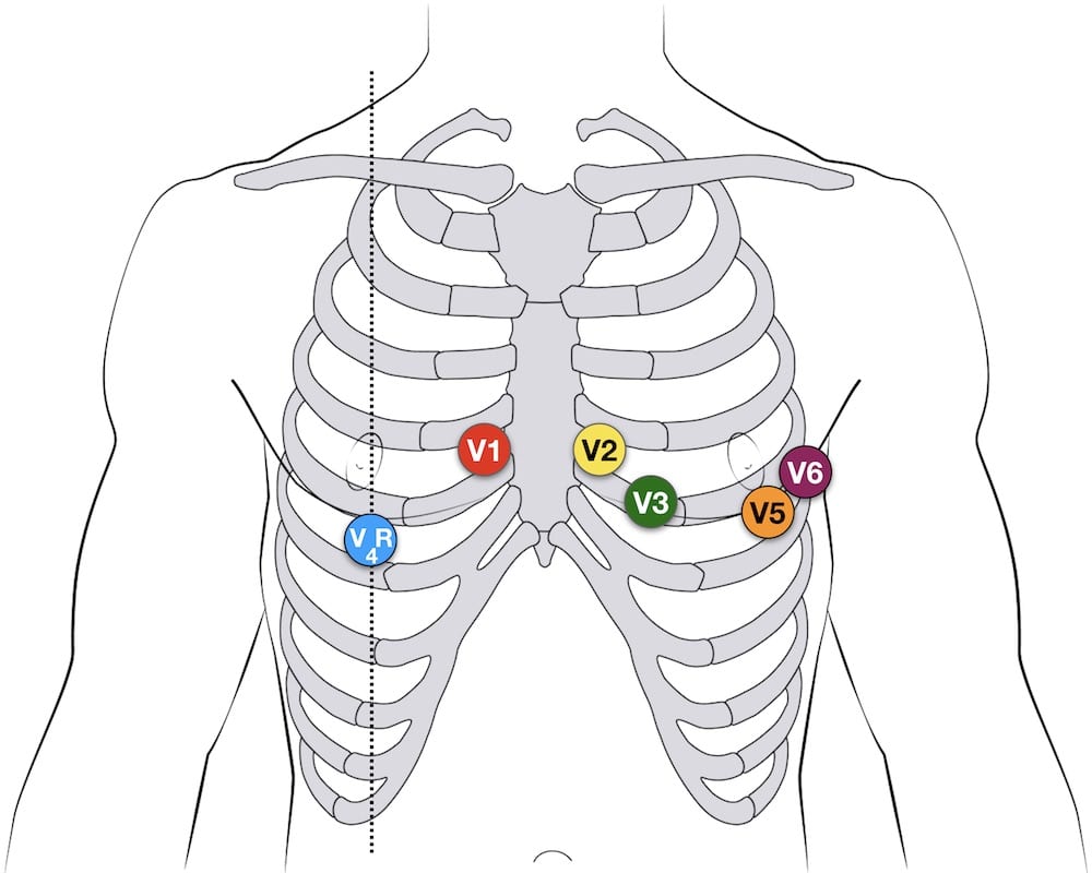
. Right sided 12 lead ECG lead placement. Red positive is referenced to white. 4th intercostal space left sternal border.
To clarify leads will equal. Placement of paediatric ECG leads. White RA Upper Right Arm Black LA Upper Left Arm Red LL Lower Left Leg 12 Lead ECG Placement.
Table 2 presents the factors which contribute successfully to the diagnosis of STEMI. V4 becomes V7 - 5th intercostal space posterior axillary line V5 becomes V8 - 5th intercostal space midscapular line V6 becomes V9 - 5th intercostal space 2 cm L of spinal column. While the 18-lead ECG is perhaps more sensitive for early detection of ischemia or infarction in practice either should be used for.
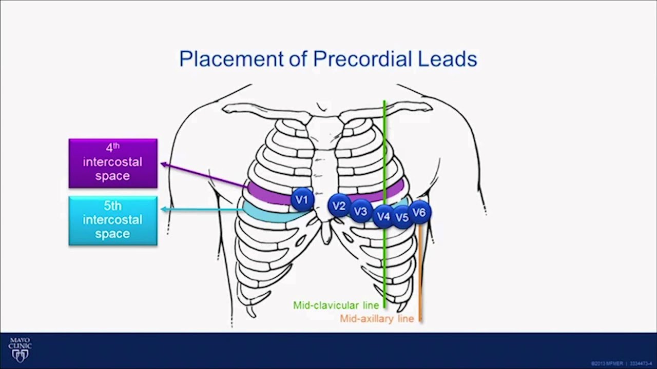
However there should be uniformity in your placement. Chest Precordial Lead Placement. V4 Placed on the midclavicular line right below the.
Aside from a 12-lead ECG placement theres something known as a 15-lead placement which includes placing leads V4-V6 on the posterior side of the patient below their left scapula see below. 4th intercostal space right sternal border. V2 Placed on the nipple line to the sternums left.
On the whole the 15-lead ECG was diagnostic of STEMI in more patients than the classic 12-lead ECG. 12-Lead ECG Interpretation Introduction This self-study package has been developed to provide a review of twelve lead interpretation as well as a review of signs and symptoms of various types of AMIs. 15 or 18 lead ECGs can be done with alternate precordial lead placement to assess for posterior- or right-sided disease.
Place the patient R lateral recumbent position to place leads. How to read an EKG The Paper The Waveform The Panl. 4th intercostal space to the right of the sternum 4th intercostal space to the leftofthe sternum directty between the V.
In young children the right ventricle normally extends to the right side of the sternum. Place limb leads on soft tissue surfaces not the bone according to the diagram on above. Here is a detailed view of the pediatric 12 Lead ECG placement approach.
For instance do not attach an electrode on the right wrist and one on the left upper arm. Watch a video on ECG leadelectrode placement. Multivariate analysis revealed that the 15-lead ECG was the sole factor significantly associated with achieving the STEMI diagnosis OR243p004 Table 2.
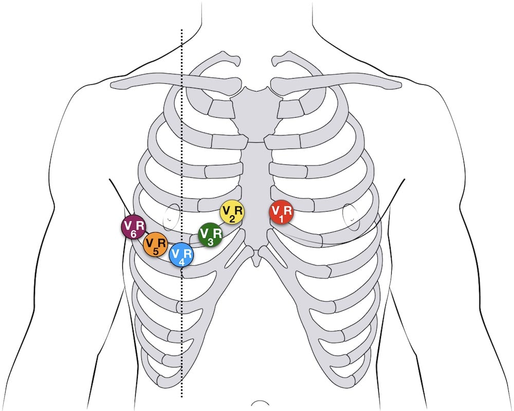
Upper leg as close to torso as possible RL green N block Above the ankle. There are several approaches to recording a right-sided ECG. A complete set of right-sided leads is obtained by placing leads V1-6 in a mirror-image position on the right side of the chest see diagram below.
12 lead placement card. It can be simpler to leave V1 and V2 in their usual positions and just transfer leads V3-6 to the right side of the chest ie. Right sided ECG electrode placement.
V1 This is placed on the nipple line to the right of the patients sternum. Place the lead at roughly the same location as you placed the lead on their right side to ensure that both arm leads give accurate readings. Chapter Page 12-Lead ECG for Acute and Critical Care Providers 2006 by Pearson Education Inc.
For female patients place leads V3-V6 under the left breast. How to read an EKG. Additional notes on 12-lead ECG Placement.
Midway between leads V2 and V4. V3 Like in grown-ups this is stationed midway right between V2 and V4. ECG limb lead placement diagram.
A 15 lead ECG is used to help diagnose a posterior and or right ventricular infarction. Pre-cordial lead placement Angle of Louis V1 What about breast tissue. 12- 15- lead ECG Section 1.
10 Lead EKG lead placement Learn with flashcards games and more for free. How to read an EKG The paper Up and down 1 box 01 mV Across 1 box 4 ms The rate 10 seconds per page. Continuing Medical Education Section 1.
12 Lead ECG Lead Placement Diagrams. To appropriately display right ventricular potentials ECGs for children in the under five-year age group must include an alternate lead V4R on the right side of the chest at a point analogous to the left-sided V4. When viewing the EKG strip V4-V6 on the strip will be referred to as V-13-15.
5th intercostal space midclavicular line. A complete set of right-sided leads is obtained by placing leads V in a mirror-image position on the right side of the chest see diagram below. The limb leads can also be placed on the upper arms and thighs.
V4V7 V5V8 and V6V9. Press down firmly on the lead to fix. 5th intercostal space anterior axillary line.

The Ultimate 12 Lead Ecg Placement Guide With Illustrations
Ecg Lead Positioning Litfl Ecg Library Basics

12 Lead Placement Guide With Diagram Video

12 15 Lead Ecg Lead Placement Youtube
Ecg Lead Positioning Litfl Ecg Library Basics
2

15 12 Lead Ecg System Anatomie Du Corps Humain Anatomie Du Corps Sante Medecine
The Ultimate 12 Lead Ecg Placement Guide With Illustrations
0 comments
Post a Comment